Showing posts with label Immunology. Show all posts

Wednesday, April 25, 2012

Study of growth property of complement C2 produced by THP-1 cells using the chemical agents –Histamine, Imidazole and β- Glycyrrhetinic acid

ABSTRACT:

Despite significant research on the role of inflammation and immunosurveillance in the immunologic microenvironment of cancer, little attention has been given to the oncogenic capabilities of the complement cascade. The complement system is the major branch of the humoral immune system.  Complement components C2 and factor B are essential for the classical and alternative pathways of complement activation, respectively, because they provide catalytic subunits for the C3 and C5 convertases, both of which are key enzymes for the complement system. Monocytes and macrophages synthesize complement proteins, thus providing an essential local source of these proteins in vivo which serve as a first-line host defense mechanism. The recent findings have shown that complement factor especially Factor B and C2 facilitates cellular proliferation and regeneration. We address this hypothesis using chemical agents Histamine, Imidazole and β- Glycyrrhetinic acid who individually influence the factor C2 production in THP-1 cells (human monocytic leukemia cell line). We investigated the growth property of complement C2 by studying the effect of these three chemical agents on the growth of THP-1 cells in time and concentration dependent manner. In this study, to our knowledge we have shown that the Complement C2 has a proliferating effect on monocytic leukemia cells. Additionally, the results show that these three chemical agents do have an influence on each other’s growth properties. Given that the traditionally held functions for the complement system include innate immunity and cancer defense, our study suggests a new way of thinking about the role of complement proteins in monocytes especially human monocytic leukemia. These results may give us leads for the treatment of monocytic leukaemias like AML-M5 which are difficult to treat.

Key words: Cancer, Complement C2, Factor B, Histamine, Imidazole and β- Glycyrrhetinic acid, THP-1, AML-M5.

For the whole article click on the link below:

https://docs.google.com/open?id=0BzdHWb7McaBDRG0tNmNFTGg2Tzg 

Tuesday, April 3, 2012

Differentiation Therapy and AML

Abstract:

Acute myelocytic leukaemia (AML) is a clonal disorder that is the consequence of acquired somatic mutations in hematopoietic progenitor cells that block normal differentiation and cell death and confer a proliferative growth advantage (Wang R. et. al., 2006). AML is basically a group of malignant bone marrow neoplasms of myeloid precursors of white blood cells.

The symptoms can be aspecific: asthenia, pallor, fever, dizziness and respiratory symptoms. Diagnostic methods include blood analysis, bone marrow aspirate for cytochemical, immunological and cytogenetical analysis, and cerebrospinal fliud (CSF) investigations. Treatment includes intensive multidrug chemotherapy and in selected cases allogenic bone marrow transplantation. Nevertheless, outcome of AML remains poor with overall survival of 35-60%. New therapeutics are required to increase the probability of cure in this serious disorder (Verschuur A. C. et. al., 2004).

Differentiation therapy has been successful as a novel treatment for APL. Several compounds including dimethyl sulfoxide, retinoic acid, phorbol ester and 1, 25-dihydroxy vitamin D3 induce AML cells to differentiate toward mature cells. Among them, retinoic acid induces AML cells to differentiate toward granulocytes, whereas 1, 25-dihydroxy vitamin D3 induces AML cells to differentiate toward monocytes (Hyun-Ock Pae a,b et. al., 2001). The following is the overview of advancement in Differentiation therapy against AML specifically AML-M5.

Key words:
AML, Hematopoietic, Symptoms, Daignostic, Therapeutics, Differentiation Therapy.

For full review click on the link below:
https://docs.google.com/open?id=0BzdHWb7McaBDM3VZMEdMS3pSdm1TSEJJbzc5UVNCUQ

Sunday, March 18, 2012

Study of growth property of complement C2 produced by THP-1 cells using the chemical agents –Histamine, Imidazole and β- Glycyrrhetinic acid

Complement System:

The complement system is the major branch of the humoral immune system. It offers a powerful defense against infection and is tightly regulated to prevent damage to self by functionally equivalent soluble and membrane regulators (Kindt T. J., 2007).

The complement system consists of a series of plasma proteins that plays an important role in host defence. There are three pathways to complement activation. The classical pathway initiates with the formation of an antibody C1q complex on the surface of a pathogen or pathogen infected cell. This complex, in turn, activates C2 via serine proteases and is itself also a serine protease. The protein C2a combines with newly cleaved protein C4a to generate a C3 convertase, C2aC4b. C3b forms the central protein complex of the complement system either by binding to complement receptors or by complexing with C2aC4b to form C5 convertase, C2aC4bC3b. This complex can bind and stabilize C5a that forms the central effector function of the complement system around which proteins C5-C9 will bind and cooperatively lyse the cell (Kindt T. J., 2007). The mannose binding pathway has a similar cascade as the classical pathway but functions independently of antibody formation. Instead, MASP1 (Mannan-binding lectin serine protease 1) and MASP2 (Mannan-binding lectin serine protease 2) binds to the mannose structures commonly found on pathogens. The Mannan-binding lectin complex is closely homologous to C1q and can activate C2 and C4. In the absence of sialic acid sugars present on normal somatic cells and which are rare on pathogens, C1q begins a lytic cascade. There is a third pathway for complement activation that begins with spontaneous activation of complement proteins. In this pathway the thioester bonds in C3 undergo hydrolysis which allows the binding of Factor B and its subsequent cleavage by plasma protease Factor D. C3b from C3 and Factor Bb from Factor B combine to form a C5 convertase (Nielsen D. G., 2009).

C3a, C4a and C5a produced acts as anaphylatoxin and regulate vasodilatation, increase permeability of blood vessels, and trigger degranulation and oxidative burst from neutrophils, eosinophiles, and basophiles. They mainly act on specific receptors to produce local inflammatory responses and when secreted in concentrations high enough to invoke a general systemic response, they cause circulatory collapse similar to an IgE mediated allergic response. They modulate the secretion of IL-6, and TNFα from B cells and serve as potent chemoattractants. C5a also works directly on neutrophils and monocytes to increase adhesion molecules, migration, and phagocytosis (Nielsen D. G., 2009).

For the whole artical click on the link below:

https://docs.google.com/open?id=0BzdHWb7McaBDSldlNUkzajBTa200Zkl0bzdKbWo2UQ


Saturday, January 21, 2012

A Study of Idiotypic Vaccine for Follicular Lymphoma Treatment


Lymphomas are a heterogeneous group of malignancies arising in the lymphoid tissue. Lymphomas are usually treated by a combination of the various therapies for eg : Chemotherapy, Radiotherapy, Bone Marrow Transplant, Stem cells procedure etc (Freedman J. ,2005). But there are lots of complications and side effects related to all the above mentioned methods (Scheinberg D. A. and Jurcic J. W., 2004).

A number of conventional therapeutic options can induce reasonably durable and sequential clinical complete responses in majority of patients (2). However, second and subsequent responses tend to be progressively shorter than those achieved earlier. In particular, most second complete responses obtained through standard chemotherapy last far less than 2 years, and the median duration is 13 months (Illidge T. and Johnson P. W. M., 2005). Follicular lymphomas (FL) are basically indolent diseases. While highly responsive to chemotherapy, the majority of lymphomas in patients remain incurable even by using various combinations of standard chemotherapy drugs. There is a need for novel therapies with less toxicity and more specific targeting of tumor cells (1).

For more on this click on link below:
A Study of Idiotypic Vaccine for Follicular Lymphoma Treatment

Wednesday, December 21, 2011

Idiotypic Vaccines for Lymphoma

Lymphomas are a heterogeneous group of malignancies arising in the lymphoid tissue. Lymphomas often start in the lymphatic system in locations such as the lymph nodes. They may then spread to other locations such as the lungs, liver or bone marrow. 25-40% of lymphomas arise at specific extranodal sites, such as splenic marginal zone B-cell lymphomas that arise in the spleen (Marcus R. and Sweetenham J. W., 2007 ) Most Lymphomas are B cell in origin, with a minority being T- cell. There are two main categories of lymphoma: Hodgkin’s Lymphoma (HL) and Non- Hodgkins Lymphoma (NHL) (Freedman J., 2005). Hodgkin’s lymphoma is a lymphoma composed of Reed-Sternberg cells surrounded by reactive non-neoplastic chronic inflammatory cells (variable numbers of lymphocytes, plasma cells, eosinophils, histocytes and neutrophils) and variable degrees of fibrosis.

Lymphoma accounts for over 3 % of cancer occurring worldwide. 75,000 people in the UK are living with lymphoma and over 13,500 people are diagnosed with lymphoma every year. The most common are B cell cancers diffuse large B cell lymphoma and Follicular lymphoma (FL) (Freedman J., 2005).

For more information click the link below:
Idiotypic Vaccines for Lymphoma

Saturday, August 13, 2011

Suicide Gene Therapy against HIV :Thymidine Kinase gene

Acquired immunodeficiency syndrome (AIDS) and its associated disorders are caused by human immunodeficiency virus (HIV). HIV exhibits a tropism for CD4+ T lymphocytes, which constitute the primary target for HIV infection in vivo. Initial infections appear to be latent and are characterized by an extended, asymptomatic stage of pathogenesis. Disease progression results in a state of prolific viral replication that eventually leads to the massive depletion of CD4+ T lymphocytes that occurs during AIDS.  Even though substantial progress has been made in the molecular characterization of HIV, therapeutic treatment of HIV mediated pathogenesis has proved difficult to attain, particularly due to uncertainties in understanding mechanisms related to the persistence of viral latency.

Resting T lymphocytes are nonpermissive for HIV replication; even then the virus efficiently binds to the CD4 receptor and is internalized.  From this latent state, the virus can be initiated into productive infection by factors that activate quiescent CD4+ lymphocytes into cellular proliferation. It appears that the latent provirus is activated by the same inducible cellular transcription factors that promote T-cell proliferation upon presentation of the appropriate antigen. This feature of HIV replication raises therapeutic possibilities; agents that are toxic to activated HIV-infected T cells would be expected to inhibit HIV replication and the ensuing pathogenesis. 

HIV-infected T cells upon activation, initiate a program of viral gene expression that is stringently controlled by two nuclear regulatory proteins coding viral genes tat and rev. The main function of tat gene product, Tat, is transcriptional activation from the viral 5' long terminal repeat (LTR) promoter by binding to structured RNA target sequence, the transactivation response element (TAR). The action of the rev gene product, Rev, is posttranscriptional; Rev selectively induces the nuclear export of a constitutively expressed pool of structural-gene mRNAs that contain a cis-acting sequence of extensive secondary structure, the Rev-responsive element (RRE). The viral cis-acting sequences TAR and RRE, whose regulatory activity is tightly controlled by Tat and Rev, respectively, afford an excellent means for the targeted expression of cytotoxic agents in HIV-infected cells that express these regulatory proteins.
 
Suicide Gene therapy is a technique for modifying the cellular genome for rendering cells sensitive to chemotherapeutics or toxins by introducing “suicide genes”. Here conditionally cytotoxic human herpes virus type- 6 thymidine kinase gene (HHV-6 TK) is used as suicide gene in Tat expressing HIV infected cells.  HSV-1 TK expression is not deleterious to mammalian cells, but it can, unlike mammalian thymidine kinase, selectively phosphorylate certain nucleoside analogs such as acyclovir and ganciclovir (GCV) to their monophosphate..  Ganciclovir(GCV),  is phosphorylated first by the viral thymidine kinase to nucleoside monophosphate (GCV-MP) and then by cell kinases to yield the triphospho form of the drug(GCV-TP).Gancyclovir triphosphate  when  incorporated into DNA, leads to inhibition of DNA synthesis .This TK-GCV system induces accumulation of p53 and increases cell surface expression of death receptors  leading to apoptosis involving the Fas-associated death domain protein (FADD) and caspases. One more advantage of the HSV-TK-GCV system is the bystander killing effect whereby HSV-TK positive cells exposed to GCV are lethal to surrounding HSV-TK negative cells via transport of GCV-MP, GCV-BP, and GCV-TP trough gap junctions to adjacent cells.Thus using HIV-2 LTR as inducible promoter for the thymidine kinase gene ,the HIV infected cells can be specifically killed .Similar strategy were  used earlier for specific killing of  HIV infected cells  where  LTR promoter is used for selective expression of  pro-apoptotic Bax gene leading to apoptotic cell death of tat expressing cells ( McCoubrie JE. et al,2004) or  LTR- herpes simplex virus (HSV) virion host shutoff gene (vhs) construct where vhs  encodes a protein which nonspecifically accelerates the degradation of mRNA molecules, leading to inhibition of protein synthesis in HIV infected cells ( Hamouda T. et al,1997).

For further information go to link stated down:
Suicide Gene Therapy against HIV : Thymidine Kinase gene

Monday, June 27, 2011

Quick Facts of IL-1R

• Interleukin-1 receptor (IL-1R) is a cytokine receptor which binds interleukin 1 (IL-1) which is a cytokine.

• There are two types of IL-1 receptor, termed type I and type II receptors, each with three extracellular immunoglobulin (Ig)-like domains but limited sequence similarity (28%) and different pharmacological characteristics.

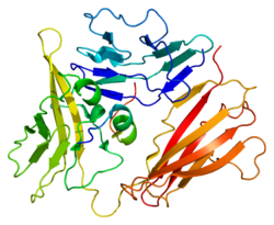
• IL-1R contains three Ig domains and, together with the highly homologous IL-1R accessory protein (IL-1RAcP), forms a receptor complex for IL-1 alpha, IL-1 beta  and IL−1 receptor antagonist (IL-1RA). Of the three Ig-like domains, domains 1 and 2 are tightly linked, while domain 3 is completely separate and connected by a flexible linker.

• Both the receptors exist in transmembrane (TM) and soluble forms: the soluble IL-1 receptor is thought to be post-translationally derived from cleavage of the extracellular portion of the membrane receptors.

• Both of them bind to the Interleukin-1 alpha, interleukin-1 beta and Interleukin-1RA which are cytokines that participate in the regulation of immune responses, inflammatory reactions, and haematopoiesis. Interleukin-1 (IL-1) exerts pleiotropic effects on a variety of tissues through binding to its receptor. interleukin-1 (IL-1R) play an important role in innate immunity by regulating the activity of distinct transcription factors such as nuclear factor-kappaB(NF-kappaB).

• IL1RB is the beta subunit of the receptor and is known also as IL1R2. The human gene encoding the beta subunit was cloned by McMahan et al (1991). In the nomenclature of CD antigens, this receptor has been given the designation CD121b.

• IL1R2 is structurally incapable of signalling and functions as a decoy receptor, binding and inhibiting the effect of IL1. IL1R2 can reduce the number of signalling complexes consisting of IL1R1, IL1RAcP, and IL1, by binding to IL1RAcP and thus has the ability to modulate the responsiveness of cells to IL1 . The type I receptor is primarily responsible for transmitting the inflammatory effects of IL-1 while type II receptors may act as a suppressor of IL-1 activity by competing for IL-1 binding.

• Also opposing the effects of IL-1 is the IL-1RA.The IL-1RA binds IL1 and transduces IL1 signals in cooperation with the co-receptor IL1RAcP to initiate an inflammatory response. In the nomenclature of CD antigens, this receptor has been given the designation CD121a. The human gene encoding the alpha subunit was been cloned by Sims et al (1989).

• All the IL-1 receptors appear to be well conserved in evolution, and map to the same chromosomal location.

• Cells may produce soluble variants of the IL1 receptors (designated sIL1R1, sIL1R2), which can compete with membrane-bound receptors for ligand binding. They can act as molecular sinks and scavengers for free ligand and can play a regulatory role by modulating ligand availability.

• IL-1 Family Receptors:

The IL-1R family consists of members that are characterized by the presence of extracellular immunoglobulin-like (Ig) domains and by an intracellular TIR domain.They are:

-IL-1ra/IL-1F3
-IL-1 Rrp2/IL-1 R6
-IL-1 RI
-IL-18 R alpha/IL-1 R5
-IL-1 RII
-IL-18 R beta/IL-1 R7
-IL-1 RAcP/IL-1 R3
-SIGIRR
-IL-1 RAPL1/IL-1 R8
-ST2/IL-1 R4
-IL-1 RAPL2/IL-1 R9


• The IL-18 receptor (IL-18R), following binding to IL-18, forms a complex with IL-18RAcP to initiate downstream signalling.

• IL1 receptor-related protein-2 (IL-1Rrp2) is the receptor for the agonists IL-1F6, IL-1F8, and IL-1F9, which also uses IL-1RAcP as a second chain. Thus, IL-1RAcP appears to be promiscuous since, in addition to IL-1RI and IL-1Rrp2, it also associates with ST2, which has recently been shown to bind IL-33.

• IL-1R2 and SIGIRR are two inhibitory receptors, the former lacks the Toll/IL-1 receptor (TIR)- domain whereas the latter contains a single Ig domain for the extracellular segment.

• The only members that still remain without any identified function in this subfamily are IL-1RAPL and TIGIRR.

• The adaptor molecules are: the Myeloid differentiation factor 88 (MyD88) and the MyD88-adaptor-like (MAL, also known as TIRAP).

• TIRAP is the TIR domain-containing adaptor protein inducing interferon-alpha.

• MyD88 has a modular structure consisting of an N-terminal death domain (DD) separated by a short linker region from the C-terminal TIR domain.

• The IL-1 receptors and TLRs (Toll-like Receptors) share a common TIR motif in their cytoplasmic domain. While IL-1 receptors respond to traditional IL-1 family ligands, the TLRs elicit inflammatory responses via the recognition of pathogen-associated molecular patterns (PAMPs) such as bacterial proteins, nucleic acids, and cell wall components.

• The TIR domain is responsible for the propagation of the signal within the cell through interaction with a complex signalling cascade. The presence of an intracellular TIR domain is the hallmark of TLR/IL-1R super family consisting of IL-1R subfamily and the TIR-domain-containing adaptor proteins.

• The IL-1 Ligands & IL-1R/TLR Family plays critical roles in inflammation and host defence. These molecules play important roles in both innate and adaptive immunity.

• MyD88 is an important signalling adaptor for both TLR and IL-1R family members. Resident skin cells utilize IL-1R/MyD88 signalling to promote neutrophil recruitment.

• A novel IL-1RI co-receptor, TILRR, links with the signalling receptor complex and enhances recruitment of the MyD88 adapter, controls induction of the Ras GTPase, and amplifies activation of NF-kappaB and inflammatory genes.

• One of the other adaptors involved are TRIF–related adaptor molecule (TRAM) where TRIF is TIR-domain-containing adapter-inducing interferon-β. TRAM contains a TIR domain in the C-terminal region and functions exclusively in the TLR4 pathway. TRAM interacts with TIR-domain-containing adapter-inducing interferon-β (TRIF) and the sterile alpha- and armadillo-motif containing protein (SARM). These adaptors bridge the TLR/IL-1R receptors to the intracellular molecules that transduce their signals into a biological response and play a central role in innate immunity.

• SARM contains a TIR domain at C-terminus, two “sterile a” motif (SAM) protein-protein interactions domains, and an Armadillo repeat motif (ARM).It functions as an inhibitor of TRIF-dependent TLR signalling.

• Another adaptor like toll-interleukin 1 receptor (TIR) domain containing adaptor protein (TIRAP) is required to act as a bridge for MyD88 in TLR2 and TLR4 signalling, while TRIF is used in TLR3 signalling and, in association with TRAM, in TLR4 signalling.

• Based on the type of adaptor molecules involved, the TLR/IL-1R-induced pathways can be sub-grouped in two classes: MyD88-dependent and MyD88-independent responses.

• In the MyD88-dependent pathway, MyD88 associates with IL-1R-associated kinases like IRAK4, IRAK1 and/or IRAK2. IRAK4 in turn phosphorylates IRAK1 and IRAK2 and promotes their association with TNF receptor associated factor-6 (TRAF6), which serves as a platform to recruit the TGF-beta activated kinase-1 (TAK1).

• Once activated, TAK1 activates the IKK complex, composed of IKK alpha, IKK beta , and NEMO (IKK gamma), which catalyzes phosphorylation and subsequent degradation of IkappaB rendering NF-kappaB free to translocate from the cytosol to the nucleus and activate NF-kappaB-dependent genes.

• TLR/IL-1R receptors associate with MyD88 through homotypic interactions between their respective TIR domains. This interaction then allows MyD88 to recruit members of the interleukin-1 receptor-associated kinase (IRAK) family (IRAK1, IRAK2, and IRAK4) through homotypic interactions between their respective Death Domains (DDs).

• The MyD88-dependent TLR/IL-1R signalling plays a vital role early in life, but becomes less important for survival during ageing. This is likely consequent to activation and/or maturation of TLR-independent innate immunity. Moreover, these findings seem to suggest that innate immunity is more important upon the very first encounter with a pathogen. Once adaptive immunity is generated, however, resistance to infection becomes quite efficient even in the absence of crucial functional components of TLR signalling.

• Once activated by their respective ligands, IL-1R, IL-18R, and TLRs engage with one or more adaptor proteins. These adaptors, namely, MyD88, MAL/TIRAP, TRIF, and TRAM are recruited, in various combinations, to the cytoplasmic domains of the receptors through homophilic interactions between Toll/IL-1 receptor (TIR) domains present in each receptor and each adaptor.

• TLR, IL-1R and TNF-R signalling to NF-kappaB converge on a common IkappaB kinase complex that phosphorylates the NF-kappaB inhibitory protein IkappaB.

• An aberrant activation of TLR/IL-1R signalling can promote the onset of inflammatory and autoimmune diseases. The development of therapeutic strategies for the control of their function have potential to cure inflammatory and immune disorders such as sepsis syndrome, asthma, atherosclerosis, Alzheimer’s disease, rheumatoid arthritis (RA).

• Pharmaceutical modulation of TLR/IL-1R signalling pathways by inhibitors, such as decoy peptides and synthetic mimetics that interfere with protein-protein interactions between signalling molecules of the TLR/IL-1R super family, might yield clinical benefits in the treatment of inflammatory and autoimmune diseases.

• The TIR domain of TLR/IL-1R proteins is a putatively suitable target. In particular, the BB-loop region may be regarded as a critical functional interface of TIR domain for its critical role in proper signalling. BB-Loop Decoy Peptides are short amino acid sequences of a protein that are expected to mimic its interaction surface and to prevent interaction of the prototype proteins with their partners.

• For e.g. A TIRAP decoy peptide consisting of the 14 amino acid-long sequence in the BB-loop (LQLRDAAPGGAIVS), fused to the Drosophila antennapedia homeodomain to facilitate the intracellular delivery, specifically blocked TLR4-induced activation of NF-kappaB without affecting the TIRAP-independent TLR9 response. In vivo administration of TIRAP inhibitory peptide counteracted the lung inflammatory response in healthy C57BL/6 mice.

• BB-loop heptapeptides derived from MyD88 and IL-18R inhibited homomeric interaction of MyD88 TIR domain or full-length MyD88 in vitro e.g. cell permeable derivative of the MyD88 BB-loop decoy heptapeptide (RDVLPGT).

• Toshchakov and colleagues performed systematic investigations of decoy cell permeable peptides containing TIR domain BB-loop sequences derived from the adaptor proteins MyD88, TIRAP, TRAM, and TRIF as well as the receptors TLR1, 2, 4, and 6. These decoy peptides were all able to inhibit, with varying activity, the TLR signalling pathways.

• Bartfai and colleagues, by focusing on TIR-domain interactions between IL-1RI and MyD88, synthesized a low-molecular-weight molecule mimetic, hydrocinnamoyl-L-valyl pyrrolidine.

• One of the most effective compounds, termed ST2825, inhibited homomeric interaction of MyD88 TIR domains. This effect was specific for TIR domains and did not affect interaction of MyD88 DD.

Although much emphasis has been placed on the development of NF-kappaB inhibitors, generic inhibition of NF-kappaB may lead to undesired side effects. Hence, a challenging objective is to develop drugs like above that block its effects in specific pathways, while leaving its physiological functions in other contexts largely intact. A promising research is going on in this line ...So watch out for this space...


Sunday, June 19, 2011

NF-κB : A Future Drug Target

NF-κB (nuclear factor kappa-light-chain-enhancer of activated B cells) is a transcription factor that started its life as a B cell-specific nuclear protein, but has matured to become one of the most extensively studied transcriptional regulators, found to control many pathophysiological processes. Its role ranges from its transcriptional regulatory functions to its participation in diverse processes, including inflammation, immunity, mucosal homeostasis and cancer. NF-kB regulates the expression of over 200 genes that are involved in a variety of functions. It is basically a protein complex found in almost all animal cell types and is involved in cellular responses to stimuli such as stress, cytokines, free radicals, ultraviolet irradiation, oxidized LDL, and bacterial or viral antigens.

NF-κB plays a key role in regulating the immune response to infection. It regulates the expression of cytokines, inducible nitric oxide synthase (iNOS), cyclo-oxgenase 2 (COX-2), growth factors, inhibitors of apoptosis and effector enzymes in response to ligation of many receptors involved in immunity including T-cell receptors (TCRs), B-cell receptors (BCRs) and members of the Toll-like receptor/IL-1 receptor super family. It also takes part in the development and the activity of a number of tissues including the central nervous system. Moreover, pathological dysregulation of NF-κB is linked to inflammatory and autoimmune diseases as well as cancer. Activation of the NF-κB/Rel transcription family, by nuclear translocation of cytoplasmic complexes, plays a central role in inflammation through its ability to induce transcription of proinflammatory genes. This pathway is activated upon appropriate cellular stimulation, most often by signals related to pathogens or stress.

In the below link, we will discuss the the regulation of NF-κB activity by IκB proteins and IκB kinase (IKK), specificity of various NF-κB proteins, their role in inflammatory disease, and the development of therapeutic strategies aimed at inhibition of NF-κB.

NF-κB A Future Drug Target.pdf